

A new process for scanning microscopic objects has been developed at Penn State. The process involves a nanosecond-pulse laser that slices microscopic objects.
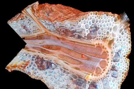
You can see the results above in a 3D model obtained from a maize root.
This is a destructive process, as the scanned object is placed on a moving platform, where the laser progressively zaps exterior layers. Each exposed layer is imaged, and these images are then combined in software to produce a full-color 3D model of the original object. But you kind of lose the object, slightly. Well, totally.
3D printing such models could be useful for teaching medical and anatomy students the interior structures of biological objects, but there could be a more fascinating use in the distant future.
Imagine a technology that could 3D print different forms of living tissue. This technology is actually being developed now, but it’s pretty crude at the moment. Eventually it could become very capable – and by having 3D models of interior structures, we would theoretically be able to print very complex biological objects.
For now, however, we can examine the scanned plant roots.
Via Kurzweil