

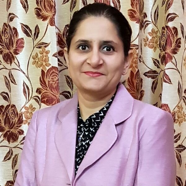
![Banu Kose [Source: Women in 3D Printing]](https://fabbaloo.com/wp-content/uploads/2020/05/image-asset_img_5eb09321495f9.jpg)
Banu Kose is a researcher and lecturer with the background in Physics Engineering (BSc), Genetics Engineering (MSc) and Biomedical Engineering (Ph.D.).
She has six years of experience with 3D anatomical modeling, 3D printing, surgical planning, medical device design, finite element analysis, and cardiovascular engineering, as well as four years of experience in teaching medical imaging physics at Istanbul Medipol University.
Nora Toure: Banu, could you let us know about your background and what brought you to 3D printing in the first place?
Banu Kose: When I was studying my MSc project, I was working on the flow simulation with patient-specific virtual artery models. I wanted to validate my simulations by creating a closed-loop circulatory system in an experimental setup by converting the virtual models into physical forms. In this process, I first involved 3D modeling and rapid prototyping. My first aim was to place replicas of arteries in this experimental setup.
Nora Toure: What was your very first experience with 3D Printing?
Banu Kose: The first things I printed were the models of two pediatric pulmonary arteries (pre-surgery and after surgery models). They were printed on an FDM printer. I had used them in a medical congress when explaining a surgical method. After I realized how much attention it was, my interest in printers became accelerated.
Nora Toure: How does 3D Printing apply to cardiovascular radiology and surgery?
Banu Kose: The treatment of complex heart disease requires a deep understanding of the 3D relationships of cardiac structures. The complexity of circulatory system disease has led researchers to search for novel imaging methods to evaluate the spatial relationships in malformed intracardiac structures.
Although the results are quite good with novel medical imaging methods of radiology, it is limited on the 2D screen of the monitor. With the image processing methods used for 3D modeling, better reconstructions are created with the existing systems of the devices. However, they cannot be sufficient in some complex cases. Through 3D printing, additional detailed information is provided for evaluating and planning the repair method.
Typically, multi-slice imaging techniques (CT, MRI, 3D Echo, etc.) are used as the source datasets to segment the anatomy to virtual preoperative models. These are the sources for 3D printed anatomical models. 3D printed cardiac models can enhance the management of patients by improving interventional and surgical planning and lead to patient-specific device designs for individual cardiac defects.
Read the rest at Women in 3D Printing


Elizabeth C. Engele (Lizzy) is a designer for social good, and a founder of MakerGirl.