
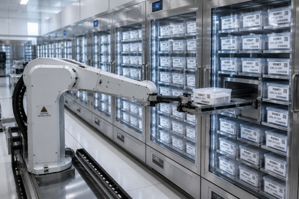
Charles G. Goulding explores how 3D printing and cryogenic preservation could transform organ transplantation by creating the world’s first biological supply chain.
The Clock Starts Ticking
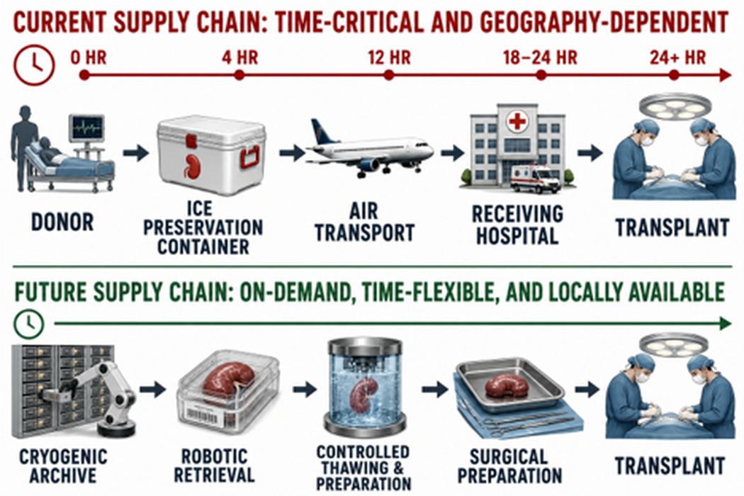
A donor kidney can save a life. It can also become a tragedy of timing. The moment an organ leaves the human body, a countdown begins. Surgeons and transplant teams work against that clock because every passing hour reduces the chance that the organ will function successfully in another patient.
This limitation shapes the entire transplant system. A donated kidney in one city may not reach the best recipient in another because biology moves faster than transportation. A patient may be too sick for immediate surgery or may need additional time for the procedure to occur safely. A rare and valuable resource can disappear simply because medicine has not yet learned how to pause it.
A world where organs could be preserved for weeks, months, or even years would follow completely different rules. Organs could be matched according to medical need rather than geography and urgency. Hospitals could maintain stores of lifesaving biological materials in the same way they maintain supplies of blood and medicine.
Researchers are beginning to build that future through cryogenic preservation. The foundation already exists in smaller forms. Fertility medicine routinely stores embryos for years. Cell therapies, including CAR-T treatments, travel through carefully controlled cryogenic supply chains between laboratories and hospitals.
The next challenge is much larger. A frozen embryo contains a small collection of cells. A human kidney contains roughly one million filtering units, an intricate network of blood vessels, and specialized structures that must survive freezing and return to function.
Why Freezing Organs Is So Difficult
These achievements demonstrate that biology can be paused. The challenge is extending that capability from individual cells and small tissues to the complex architecture of an entire organ.
A kidney contains approximately one million nephrons. Each nephron contains specialized cells and tiny blood vessels responsible for filtering blood and maintaining the body’s chemical balance. Every organ is a landscape of microscopic passages, membranes, and cellular communities. Successful preservation requires protecting that landscape as a complete system.
The difficulty grows with size. The exterior of an organ cools and warms differently than deeper regions. Water shifts between cells. Different materials contract at different rates. Temperature gradients create mechanical stresses capable of damaging delicate biological structures.
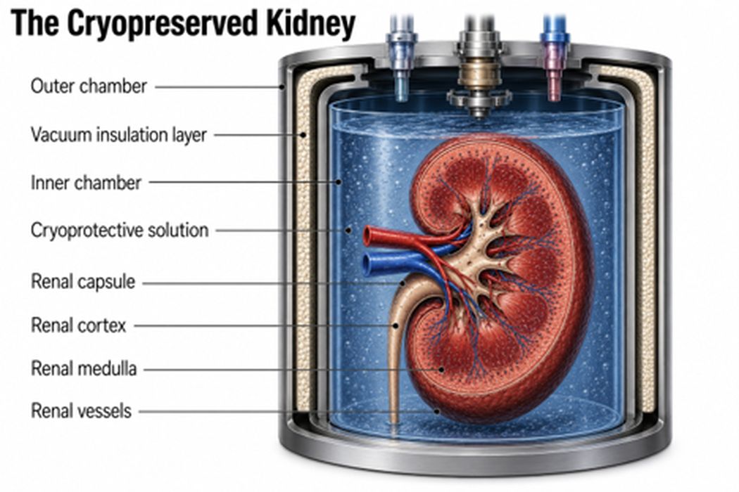
Successful cryopreservation depends on controlling chemistry, temperature, and mechanical forces across an entire organ.
Vitrification: Preserving Life Without Ice
Vitrification represents one of the most promising approaches to long-term organ preservation. Scientists introduce cryoprotective agents, often called CPAs, that replace portions of the water inside tissues and allow biological material to enter a glass-like state. The goal is not simply to make an organ cold. The goal is to control what happens inside every microscopic corner of a three-dimensional structure.
That requirement exposes one of the central challenges of cryogenic medicine. A kidney is not a solid block of tissue. It contains millions of microscopic structures, narrow blood vessels, and complex pathways that must all receive the right concentration of protective chemicals. Too little cryoprotective solution allows damaging ice to form. Too much can injure living cells.
This is where the logic of 3D printing begins to enter the story. Engineers can use additive manufacturing to rethink the internal architecture of biological structures. A printed tissue can contain vascular channels, pores, and pathways designed to move fluids more effectively. The same structures that eventually carry nutrients and oxygen inside the body may also help distribute cryoprotective agents during preservation.
Freezing is only half of the challenge. A successful organ must also return safely to normal temperatures. Uneven warming can create fractures. A process known as devitrification can trigger the formation of damaging ice crystals as tissue leaves its glass-like state.
Researchers at the University of Minnesota have explored nano-warming as a solution. Magnetic nanoparticles placed throughout biological materials can be activated by radiofrequency energy, generating heat from inside the tissue rather than relying entirely on heat moving inward from the surface.
The same principle appears repeatedly throughout the future of cryogenic medicine: the more control scientists have over the internal environment of an organ, the greater their ability to preserve it. 3D printing offers a powerful new way to create that control by allowing engineers to design the pathways through which chemicals, fluids, and eventually heat can move.
3D Printed Capillaries
A human body contains approximately 60,000 miles of blood vessels. The smallest capillaries measure only a few micrometers in diameter. These microscopic pathways deliver oxygen, nutrients, hormones, and immune signals to nearly every cell.
Recreating those networks remains one of the central engineering challenges in tissue manufacturing.
At Harvard University’s Wyss Institute, Professor Jennifer Lewis and her team developed sacrificial writing into functional tissue, commonly known as SWIFT. The process prints a temporary material through a dense collection of living cells. The temporary material is removed, leaving hollow channels capable of carrying fluids through the tissue.
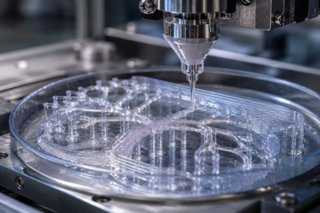
The work addressed one of the greatest challenges in creating thick engineered tissues. Cells located far from a nutrient source struggle to survive. A printed vascular network creates pathways through which fluids can circulate.
Lewis has identified vascularization as a critical bottleneck in creating clinically useful tissues. The ability to organize channels throughout a biological structure gives engineers control over the internal geography of living material.
That control carries major implications for cryogenic preservation. Cryoprotective agents must move throughout an organ before freezing and then be carefully removed during warming. A printed network of channels can serve as a transportation system for those fluids.
Engineers can modify channel diameter, branching patterns, and spacing through digital design. A scaffold can contain pores that reduce thermal stress. A tissue can contain regions optimized for fluid movement during preservation.
The architecture of an organ becomes another variable engineers can adjust.
Designing Organs for Storage
A donated organ arrives with a fixed design. Every blood vessel, membrane, and microscopic structure developed through human biology. A printed organ begins as a digital file. That difference may become one of the most important advantages in the future of cryogenic medicine.
The first major step was proving that living tissue could be manufactured at all. Organovo became one of the earliest companies to commercialize three-dimensional printed human tissues. Its printed liver models gave pharmaceutical companies more realistic systems for studying drug toxicity. More importantly, Organovo showed that a biological structure could be produced repeatedly with a known architecture and consistent characteristics.
Once tissues become manufacturable, engineers can begin asking a new question: what should the ideal design look like?
BICO and its CELLINK division provide researchers with bioprinting platforms capable of depositing hydrogels containing living cells in precise arrangements. A scientist can alter the spacing between cells, the composition of supporting materials, or the geometry of the surrounding structure. Each change creates another version of the biological blueprint that can be tested and improved.
Aspect Biosystems has pushed this level of control further through microfluidic bioprinting. Multiple streams of cells and biomaterials can be positioned with remarkable precision, allowing researchers to build increasingly complex tissues with controlled internal organization.
The importance of this precision extends far beyond making a tissue function inside the human body. A future printed organ will also need to survive manufacturing, transportation, storage, freezing, and warming. The pathways that carry nutrients may also carry cryoprotective agents. The scaffold supporting cells may also absorb mechanical stresses created by extreme temperature changes.
Engineers have spent centuries designing products around the conditions they will experience after leaving the factory. A future biological product may follow the same principle. Its architecture may be shaped not only by what the body needs, but also by what the supply chain demands.
A digital design can be modified repeatedly. A channel can become wider. A hydrogel can become stronger. A cellular arrangement can be adjusted to improve survival after preservation. The next version can be printed, tested, and refined.
| Biological Structure | Why It Matters | Current State of 3D Printing | Representative Companies/Projects | Cryogenic Relevance |
| Skin | Burns, wounds, and reconstructive surgery | One of the most advanced applications, with printed skin grafts and tissue models advancing toward clinical use | 3DBio Therapeutics; Wake Forest Institute for Regenerative Medicine | Stored skin grafts could provide rapid access for hospitals treating severe injuries |
| Cartilage | Joint injuries and facial reconstruction | A strong early target because of its relatively simple architecture and absence of blood vessels | 3DBio Therapeutics; university research programs | Long-term storage could create inventories of personalized implants |
| Liver tissue | Drug testing, disease modeling, and future transplantation | Functional liver tissues exist, while a complete transplantable liver remains a major engineering challenge | Organovo; CELLINK-enabled research laboratories | Preservation is essential for moving from laboratory production to widespread distribution |
| Blood vessels | Provide nutrients and oxygen throughout larger tissues and organs | Major advances have occurred in printed vascular channels and branching networks | Harvard Wyss Institute; Jennifer Lewis’s vascular printing research | Internal channels may improve delivery and removal of cryoprotective agents |
| Heart tissue | Heart failure, cardiac damage, and regenerative repair | Printed patches and smaller structures have demonstrated promise, while complete hearts remain a long-term goal | Tel Aviv University cardiac bioprinting research; academic laboratories | Storage could allow engineered cardiac tissues to be available when patients need them |
| Kidney | One of the largest areas of unmet transplant need | Researchers have created kidney tissues and organoids; a complete transplant-ready kidney remains a major frontier | Academic bioprinting programs; regenerative medicine companies | Successful preservation could transform transplant logistics |
| Lung tissue | Respiratory failure and donor shortages | Airway structures and lung tissue models are advancing; complete lungs remain among the most difficult goals | United Therapeutics and regenerative medicine initiatives | Complex air sacs and vascular structures create extraordinary preservation challenges |
The First Biological Supply Chain
With a biological supply chain, a surgeon in a community hospital can open a cryogenic storage unit and remove a tissue that was manufactured months earlier in a specialized facility hundreds of miles away. The tissue arrives with a digital record describing its design, the cells used to create it, the conditions under which it was printed, and every stage of its preservation journey.
That scenario begins long before the tissue enters a freezer. It begins with a digital blueprint. A researcher using a CELLINK bioprinter can adjust the spacing of vascular channels, alter the composition of a hydrogel, or change the arrangement of living cells before a single layer is printed. A design that performs poorly during preservation can be modified. The next version can be produced and tested.
Aspect Biosystems extends that level of control through microfluidic bioprinting. Its technology places multiple biomaterials and cell populations into precisely organized structures. Those design choices influence how a tissue functions inside the body and how successfully it tolerates freezing, storage, and warming.
This ability to repeatedly manufacture and improve living structures represents a fundamental shift. Organovo helped establish this idea by producing printed liver tissues with consistent architecture for pharmaceutical research. The achievement was not simply a better laboratory model. It demonstrated that living tissue could become a manufactured product with defined specifications.
Can Companies Claim the R&D Tax Credit for Bioprinting and Organ Cryopreservation Innovations?
Yes. Life sciences companies, medical device manufacturers, and regenerative medicine firms can claim the Section 41 Research and Development (R&D) Tax Credit for Qualified Research Expenses (QREs) incurred while designing, prototyping, and validating advanced bioprinting workflows, specialized bio-inks, and cryogenic preservation methodologies.
How Do Bioprinting Workflows and Internal Vascular Design Qualify under Section 41?
Developing manufactured living tissues introduces extreme technical uncertainty regarding cell viability, nutrient delivery, and structural integrity. When conventional tissue scaffolding fails to prevent cellular necrosis in deep regions, the engineering hours, iterative CAD modeling, and biomaterial inputs used to design proprietary vascular networks qualify as QREs.
R&D Case Study: Sacrificial Tissue Manufacturing
- Industry: Regenerative Medicine & Biomanufacturing
- Innovation: Development of Sacrificial Writing Into Functional Tissue (SWIFT), utilizing microfluidic bioprinting platforms to extrude temporary sacrificial channels through dense cell matrices, which are subsequently removed to leave open, fluid-carrying conduits.
- Impact: Eliminated technical uncertainty surrounding vascularization bottlenecks in thick engineered tissues, establishing scalable, fluidic pathways capable of distributing cryoprotective solutions and surviving devitrification stresses.
Does Cryogenic Preservation and Nano-warming Development Qualify for Tax Incentives?
Yes. While freezing biology is a logistical step, developing the precise chemistry, thermal profiles, and hardware to successfully pause and revive complex organ architectures represents an eligible process of experimentation.
| Activity Category | Qualifying R&D Sub-Component | Tax Treatment (Section 41 vs. Section 174A) |
| Biomaterial Formulation | Designing and testing precise concentrations of Cryoprotective Agents (CPAs) to induce vitrification without causing cellular toxicity. | Section 41: Eligible for immediate tax credit via employee hours and specialized chemical consumption during lab trials. |
| Thermal Engineering | Researching magnetic nanoparticle distribution and radiofrequency energy activation to achieve uniform nano-warming throughout frozen tissues. | Section 41: Eligible where iterative physical testing is required to eliminate mechanical stress fractures during thawing. |
| Digital Bio-Blueprint Design | Adjusting cell spacing, hydrogel tensile strength, and pore geometry inside CELLINK or Aspect Biosystems bioprinters to withstand extreme temperature transitions. | Section 174A: Advanced digital modeling, simulation software setups, and initial CAD-blueprint programming. |
Conclusion
Every major manufacturing industry depends on the separation between production and use. A factory builds products before customers need them. Warehouses maintain inventory. Quality systems document how each product was created.
Biological manufacturing is moving toward the same model. A specialized facility could print batches of skin grafts, cartilage, vascular tissues, and organ components. Each product could undergo testing, enter cryogenic storage, and remain available until a physician requires it.
The transition would be especially powerful for patients far from major research hospitals. A therapy created in a centralized manufacturing center could reach a smaller hospital with the same detailed production history as a high-value industrial component.
With 3D printing, a digital design can be improved, reproduced, stored, and distributed with a level of control that natural donation has never allowed.
Blood banks changed medicine by separating the moment blood was donated from the moment a patient needed it. Cryogenic biomanufacturing can create the same separation for engineered tissues.
The first successful biological supply chain will accomplish something profound: allow living tissues to wait.