
Researchers at Weill Cornell Medicine and Cornell Engineering offer the promise of grafts with well-defined anatomy and the correct biomechanical properties.
Researchers at Weill Cornell Medicine and Cornell Engineering have assembled a replica of an adult human ear that reportedly looks and feels natural, thanks to state-of-the-art tissue engineering and a 3D printing. The study, published online in Acta Biomaterialia, offers the promise of grafts with well-defined anatomy and the correct biomechanical properties for those who are born with a congenital malformation or who lose an ear later in life.
“Ear reconstruction requires multiple surgeries and an incredible amount of artistry and finesse. This new technology may eventually provide an option that feels real for thousands needing surgery to correct outer ear deformities,” said Dr. Jason Spector, Chief of the Division of Plastic and Reconstructive Surgery at NewYork-Presbyterian/Weill Cornell Medical Center and a professor of surgery (plastic surgery) at Weill Cornell Medicine.
Many surgeons build a replacement ear using cartilage removed from a child’s ribs, in an operation that can be painful and scarring. Although the resulting graft can be crafted to resemble the recipient’s other ear, it generally does not have the same flexibility.
One way to produce a more natural replacement ear is to enlist the aid of chondrocytes, the cells that build cartilage. In earlier studies, Dr. Spector and his colleagues used animal-derived chondrocytes to seed a scaffold made of collagen, a key component of cartilage. Though these grafts developed successfully at first, over time the well-defined topography of the ear – its familiar ridges, curves, and whorls – was lost. “Because the cells tug on the woven matrix of proteins as they labor, the ear contracted and shrank by half,” said Dr. Spector.
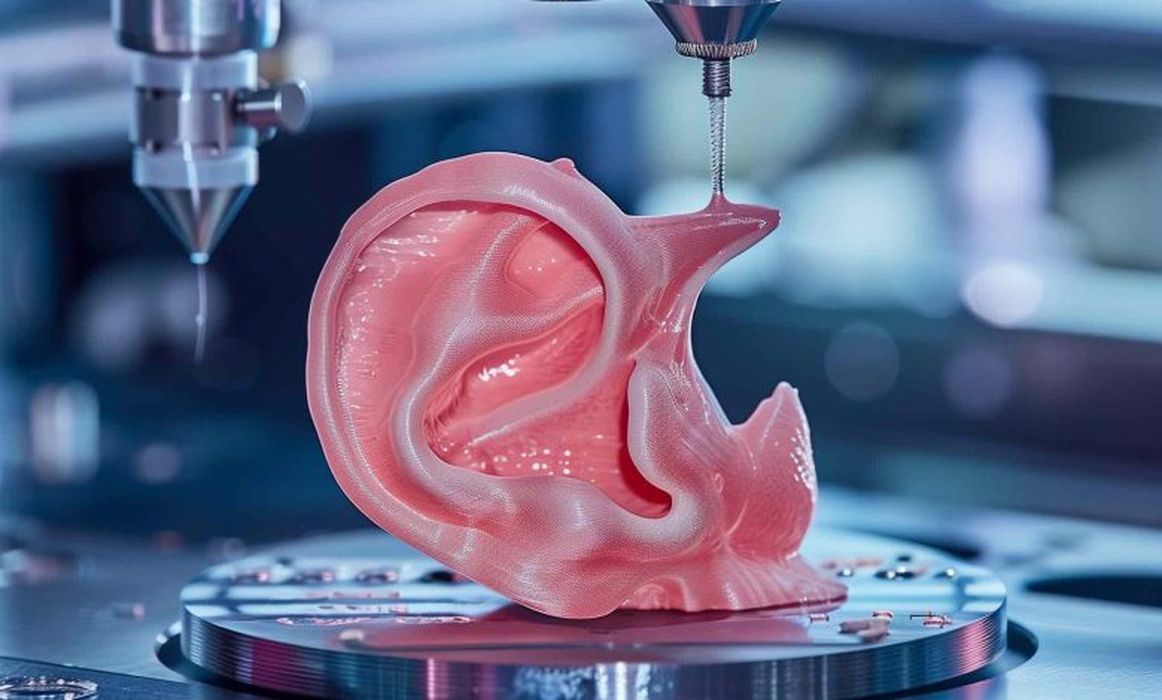
To address this problem in this study, Dr. Spector and his team used sterilized animal-derived cartilage treated to remove anything that could trigger immune rejection. This was loaded into intricate, ear-shaped plastic scaffolds that were created on a 3D printer based on data from a person’s ear. The small pieces of cartilage act as internal reinforcements to induce new tissue formation within the scaffold. Much like rebar, it strengthens the graft and prevents contraction.
Over the next three to six months, the structure developed into cartilage containing tissue that closely replicated the ear’s anatomical features, including the helical rim, the ‘anti-helix’ rim-inside-the-rim, and the central, conchal bowl. “That’s something that we had not achieved before,” said Dr. Spector.’
Read the rest of this story at VoxelMatters