
Two photon polymerization has now moved from “on cells” to “inside cells”, opening a strange, new frontier.
Two photon polymerization (TPP) is one of the sharpest tools in microfabrication, today routinely used to produce micro optics, photonic components, and biomedical scaffolds with features well below a micron. The technique relies on a femtosecond laser to trigger polymerization only at the focal voxel, which is why it can produce intricate free standing structures without the usual layer wise constraints seen in larger format additive processes.
Biocompatible TPP has mostly lived outside cells: printing scaffolds for cell growth, printing hydrogels with embedded cells, or printing structures in tissue like environments. There have even been demonstrations of “in vivo like” printing inside small living organisms, and at least one report of printing inside a synthetic cell. But actual fabrication inside a living mammalian cell has not been done, largely because you need a photoresist that a cell can tolerate both before and after polymerization.
That is the gap addressed by researchers at the J. Stefan Institute and University of Ljubljana, who report direct 3D printing of microstructures inside living HeLa cells. Their key trick is to inject a tiny droplet of photoresist into the cell, then polymerize only the parts you want, and let the rest dissolve away.
Printing In Cytosol With A Commercial TPP Platform
The team microinjected a negative tone photoresist droplet into live HeLa cells, then used a commercial Nanoscribe system (a Photonic Professional GT2) with a 780nm femtosecond laser to write a designed pattern inside the droplet. Because near infrared light passes through the cell and medium with minimal absorption, polymerization occurs only at the intense focal spot, producing a solid microstructure while leaving surrounding cytoplasm basically untouched.
Material choice was the make or break step. The authors selected Nanoscribe IP S because it seems to be relatively well tolerated by cells even before polymerization, and it is slightly water soluble so unpolymerized resin can slowly dissolve. That solubility means there is a practical time window: the operators typically had one to two hours after injection to complete printing, depending on droplet size.
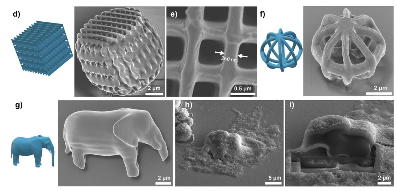
Within droplets about ten to fifteen microns wide, they printed objects around ten microns in scale, including a tiny 10 µm elephant, multiple separate structures inside one cell, and even simple logos. The actual laser writing is fast: a ten micron class object took roughly three to ten seconds to fabricate, depending on volume. At this scale, resin viscosity and short exposure times meant the structure did not drift during printing, so they did not need supports.
Resolution, Distortion, And The Reality Of Droplet Optics
Printing inside a droplet embedded in cytoplasm sounds like an optical nightmare, because the refractive index mismatch can shift focus and blur the voxel. IP S in liquid form is reported around 1.48, while cytoplasm is closer to 1.36 to 1.39, so refraction at the droplet boundary is unavoidable.
The team modeled the effect with ray optics simulations and found the focal displacement stays small, reaching about 0.5 µm near the droplet edge. Defocusing remained below a 400 nm diffraction limit in more than ninety percent of the droplet volume, implying most of the printable region behaves close to “bulk photoresist” conditions.
They backed this up with scanning electron microscopy of prints made in droplets (outside cells but in a refractive index matched solution). A woodpile style test structure showed walls as thin as 260nm on an approximately 0.8 µm grid. Cross sections of an elephant printed inside a cell showed a dense structure without obvious porosity at the 100nm scale.
Mechanically, the printed IP S polymer is stiff for a cell environment, with a Young’s modulus around 2.1GPa, so even thin features stayed rigid during cell migration and division.
Cell Health, Division Timing, And The Cost Of Intracellular Manufacturing
Printing a solid object into a living cell is never going to be “free”, and the study does not pretend otherwise. The authors observed that cells containing large printed objects showed deformed internal organization, with the nucleus reshaping around the structure. That is not surprising, but it is important: this approach can intentionally modify cell mechanics by adding a rigid inclusion where biology did not plan for one.
When they quantified division timing, cells carrying large printed structures (greater than about five microns) took longer to divide, typically delayed by about one hour and sometimes by several hours. Cells with small printed structures (under about three microns) behaved more like controls. In other words, intracellular printing can shift phenotype by brute force geometry alone.
Viability numbers were not great. After twenty four hours, non manipulated controls showed about ten percent non viable cells under their typical handling conditions. Microinjection alone raised non viability to around fifty percent, silicone oil droplet injection landed around forty four percent, and the printed structure condition was about fifty five percent. The message is clear: membrane penetration and the mechanics of adding a droplet are likely the dominant damage sources, while resin toxicity and illumination may be secondary contributors.
It’s clear there are many challenges with this approach, yet the researchers were able to successfully 3D print inside living cells. At least the ones that lived, that is.
Via Wiley