
Charles R. Goulding and Preeti Sulibhavi unpack how AI-driven design and additive manufacturing are changing how surgeons think about fit, fusion, and recovery.
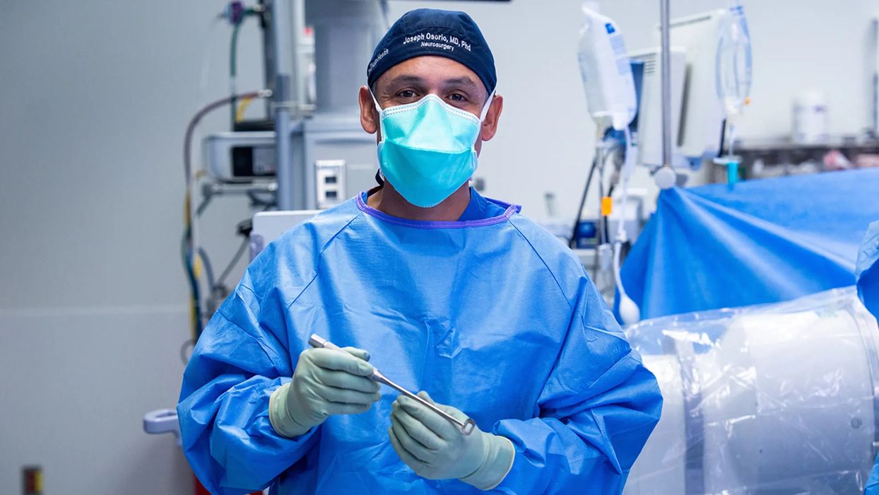
On July 14 2025, neurosurgeon Dr. Joseph Osorio and his team at UC San Diego Health performed a landmark operation — the first anterior cervical spine surgery using a fully personalized implant tailored specifically to a patient’s anatomy using advanced imaging, AI-assisted planning, and 3D printing.
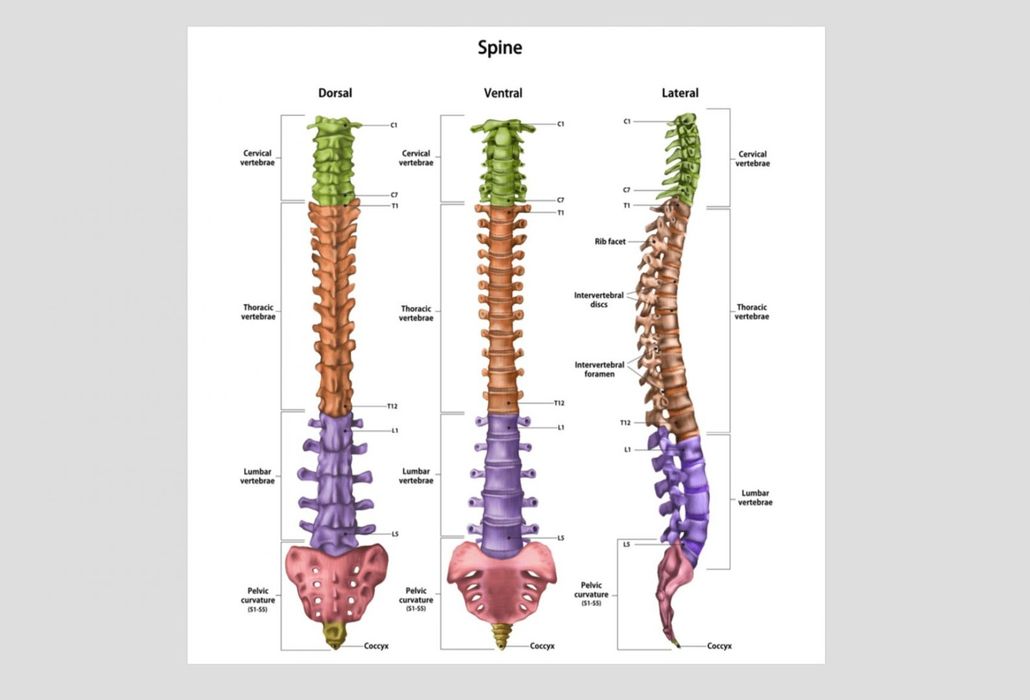
Anterior cervical fusion — the procedure used in this case — is one of the most common spine operations performed worldwide. Traditionally, it involves an incision in the front of the neck, removal of a damaged intervertebral disc, and placement of a synthetic spacer or cage between the vertebrae to support fusion and maintain alignment. Surgeons then secure the construct with screws or plates to promote stability and bone healing.
From One-Size-Fits-All to One-Person-Only
For decades, the implants used in anterior cervical surgery have been “off-the-shelf” devices manufactured in standard sizes and shapes. Surgeons select from a catalogue of generic cages and plates that approximate the patient’s anatomy. While this approach works well in many cases, it still requires the patient’s body to adapt to an implant that wasn’t designed specifically for them. This can affect the precision of spinal alignment, the interface between bone and implant, and ultimately the biomechanics of the cervical spine.
In contrast, the UC San Diego case used technology that models and matches the patient’s spine like a fingerprint — a “one patient, one implant” philosophy. “Every spine is unique, just like a fingerprint,” Dr. Osorio said. “With this technology, we can create an implant specifically for each patient, instead of asking their body to adapt to a standard device.” This shift — from approximate fit to precise fit — is more than semantics. It reflects a deeper understanding of cervical spine biomechanics, where even small misalignments can alter load distribution, motion, and long-term stability.
What’s Different About This Surgery?
From a neurosurgeon’s perspective, the most striking differences between this custom approach and traditional surgery are in preoperative planning, implant design, and intraoperative fit:
1. Detailed Anatomical Mapping with Advanced Scans.
Instead of relying on general implant dimensions, the surgical team started with high-resolution imaging (likely CT and/or MRI) to capture the patient’s cervical vertebral contours and spatial relationships in precise 3D. These scans form the foundation of both digital design and intraoperative execution.
2. AI-Assisted Design.
Artificial intelligence was used to analyze the imaging data and determine the optimal implant geometry. Rather than manually sketching or templating the design, AI algorithms can rapidly evaluate the exact shape, curvature, height, and footprint needed to match the vertebral endplates and maintain physiologic alignment. This includes adjustments for lordosis (natural cervical curvature), disc height restoration, and pathological features like deformities or asymmetries.
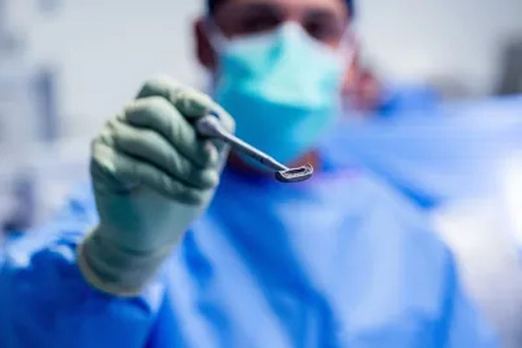
The implant was then 3D printed from medical-grade titanium, a material chosen for its strength, biocompatibility, and track record in orthopedics.
3. Push-Button Personalization vs. Manual Sizing.
Surgeons using traditional implants have to compromise — selecting the nearest size from discrete options and modifying placement intraoperatively to achieve the best fit. With custom implants, there’s no “closest match.” The implant is digitally sculpted to match the patient’s vertebrae and spatial orientation before ever entering the operating room.
4. Operating Room Efficiency and Fit.
Because the custom device precisely matches the patient’s vertebral anatomy, intraoperative adjustments can be minimized. This potentially shortens surgical time and reduces the guesswork around implant placement, screw trajectories, and bone preparation. That’s a meaningful advantage in a region as delicate as the cervical spine.
Spine Like a Fingerprint: Why Personalization Matters
Every human spine differs in curvature, bone density, and segmental proportions. Think of fingerprints — no two are alike. The same is true for cervical vertebrae: they vary in width, height, angle, and the relative geometry between adjacent levels. Traditional implants work on the assumption that a standardized design can approximate this complex variability. But when implants are custom-designed using data from a patient’s own anatomy, the fit becomes exact and individualized — and that changes the biomechanics.
From a neurosurgical standpoint, this has several implications:
- Optimal Alignment: Custom implants can restore the natural curvature of the cervical spine more precisely, which is critical for long-term function and adjacent segment health. Misalignment can accelerate degeneration above or below the fused level.
- Bone-Implant Contact: Better contact between bone and implant can lead to more uniform load transfer, reducing stress risers and promoting healthier fusion biology.
- Decreased Micromotion: Custom fit limits micromotion at the implant-bone interface, which can reduce pain and improve stability while the bone heals.
3D Printing Medical-Grade Titanium: How It Works
The implant in this case was additively manufactured — 3D printed using medical-grade titanium — but the specifics of the machine and technique aren’t public in the UC San Diego press release itself. Typically, implants like this might be produced using laser powder bed fusion or electron beam melting (EBM), both of which are FDA-recognized methods for titanium orthopedic implants. These technologies allow highly controlled fabrication of complex geometries from titanium powder, layer by layer, with tight tolerances.
Why titanium? Because it:
- Is biocompatible and well-tolerated by human tissue.
- Has the strength to support spinal loads.
- Can be printed with porous lattice structures that encourage bone ingrowth.
- Doesn’t corrode in the body.
In additive manufacturing, the digital implant design from the AI system is converted into a set of machine instructions for the 3D printer. The printer then constructs the implant layer by micrometer-thin layer, fusing titanium powder using lasers or electron beams under a controlled atmosphere. Post-processing includes heat treatment, cleaning, and sterilization before surgical use.
AI’s Role in Implant Design
Artificial intelligence accelerates and refines the planning phase. Rather than manual templating, AI can interpret 3D anatomy and propose an optimized implant shape that:
- Matches the patient’s vertebral endplates.
- Maintains correct spinal curvature.
- Predicts the best locations for fixation features.
- Minimizes areas of high stress.
This is far beyond simply “choosing size and shape” — it’s a data-driven custom geometry generated for unique anatomy and pathology.
Why The Joint Commission Recognition Matters
UC San Diego Health’s spine program has earned accreditation from The Joint Commission for excellence in spinal surgeries, a distinction that signals quality and safety.
The Joint Commission’s Advanced Certification in Spine Surgery (ACSS) emphasizes rigorous standards to minimize complications, ensure evidence-based practice throughout the care continuum, and enhance outcomes. Programs with this certification must:
- Adhere to clinical practice guidelines.
- Collect and report performance measures such as infection rates, neurological outcomes, and patient-reported outcomes.
- Demonstrate multidisciplinary coordination from preoperative assessment through postoperative recovery.
For patients, this certification means they’re being treated in a program that meets a high bar of clinical performance and continuous quality improvement, not just a technological showcase.
Benefits for Post-Op Recovery and Long-Term Outcomes
One of the most significant promises of this technology is its potential to improve recovery and function:
1. Faster Recovery and Less Pain:
When an implant fits precisely, it can reduce soft-tissue disruption and promote earlier biomechanical stability, potentially translating to less pain and earlier mobility.
2. Better Fusion Biology:
Exact fit means more surface contact between bone and implant, which may promote bone ingrowth and successful fusion, a key factor in long-term success.
3. Lower Rates of Revision Surgery:
Misfit and suboptimal alignment are among the reasons patients need revision surgeries. By tailoring the device to the individual, there’s potential to lower long-term revision rates.
4. A More “Natural” Integration:
Custom geometry may allow the implant to behave more like native bone in how it distributes load, which could translate into better functional biomechanics over time.
The Research & Development Tax Credit
The now permanent Research & Development Tax Credit (R&D) Tax Credit is available for companies developing new or improved products, processes and/or software.
3D printing can help boost a company’s R&D Tax Credits. Wages for technical employees creating, testing and revising 3D printed prototypes can be included as a percentage of eligible time spent for the R&D Tax Credit. Similarly, when used as a method of improving a process, time spent integrating 3D printing hardware and software counts as an eligible activity. Lastly, when used for modeling and preproduction, the costs of filaments consumed during the development process may also be recovered.
Whether it is used for creating and testing prototypes or for final production, 3D printing is a strong indicator that R&D-eligible activities are taking place. Companies implementing this technology at any point should consider taking advantage of R&D Tax Credits.
Looking Ahead
This first cervical case represents more than a technical milestone. For neurosurgeons, it’s evidence of what’s possible when precision imaging, AI design, and additive manufacturing converge. Dr. Osorio envisions a future where every implant — from spine to hip to knee — is made for one person, not mass-produced for everyone.
This integration of personalized data and custom fabrication could reshape how we think about implants across surgical disciplines. For patients, it means implants that feel and function more like a natural extension of their anatomy — engineered with intention rather than approximation.