
Anthony Palumbo and Charles Goulding examine how additive manufacturing is accelerating embryo screening and IVF innovation by enabling high-resolution microfluidic lab-on-a-chip platforms, biocompatible culture scaffolds, and next-generation cryopreservation and micromanipulation tools.
Introduction
The fields of additive manufacturing (AM) and reproductive medicine are converging in unexpected ways. As in vitro fertilization (IVF) laboratories adopt high-tech methods for embryo screening – including advanced genetic tests like polygenic risk assessment – they increasingly rely on custom microdevices and tools. 3D printing is emerging as a key enabler, allowing rapid prototyping and fabrication of specialized lab-on-a-chip devices, biocompatible scaffolds, and precision lab equipment. These innovations aim to improve embryo handling, culture conditions, and cryopreservation, ultimately boosting IVF success rates. This article explores the technological intersections between AM and embryo screening, highlighting how 3D printing is used to manufacture lab-on-a-chip systems, enable bioprinting for cell handling, create new cryopreservation tools, and enhance IVF lab equipment. Each example is backed by research or case studies, illustrating a technical and analytical view of this fast-evolving domain.
3D Printed Lab-on-a-Chip Devices for IVF and Genetic Screening
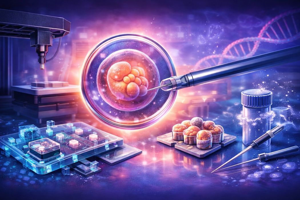
One of the most promising applications of AM in reproductive medicine is the fabrication of microfluidic lab-on-a-chip devices for IVF. These devices miniaturize and automate processes such as fertilization, embryo culture, and genetic testing. Traditionally, microfluidic chips were fabricated using etching or molding, but high-resolution 3D printing, particularly two-photon polymerization (2PP), now enables rapid production of complex microchannels and chambers with feature sizes down to tens of microns.
A leading example is the work of Fertilis in collaboration with UpNano GmbH. Fertilis developed a 3D printed microfluidic embryo culture device designed to replicate physiological conditions found in vivo. The transparent chip contains microchannels and chambers allowing embryos to be immobilized while precisely controlling media flow and environmental conditions. By eliminating repeated manual transfers between petri dishes, the system reduces mechanical stress on embryos and improves consistency across culture stages. Clinical modeling suggests that this approach may reduce the number of implantation cycles required per successful pregnancy by approximately 30–40%.
The technical achievement behind this IVF lab-on-a-chip was enabled by 2PP 3D printing using a special photopolymer resin called UpFlow. UpNano’s NanoOne 2PP printer can print sub-micron features, and the UpFlow material has very low viscosity before final curing, which allows thorough flushing of uncured resin from hair-thin channels. The low viscosity allows uncured resin to be fully flushed from channels smaller than 50 µm, overcoming a longstanding manufacturing limitation in printed microfluidics. The material also provides high optical transparency and low autofluorescence, which is critical for embryo observation under fluorescence and optical microscopy. The shift to additive manufacturing reduced production time from approximately two weeks to roughly four hours, enabling faster iteration and scalable manufacturing.
Beyond embryo culture, AM-fabricated lab-on-a-chip devices also support genetic screening workflows. As IVF clinics expand from single-gene preimplantation genetic testing to polygenic screening, efficient handling of extremely small DNA samples becomes increasingly important. Microfluidic biochips fabricated using 3D printing can integrate on-chip DNA amplification, high-throughput PCR, and fluorescence-based assays for chromosomal or gene-level analysis.
For instance, Pixelbio in Germany used stereolithography 3D printing to fabricate molecular biochips for multiplex genetic detection. Their printed chips integrate microchannels and reaction chambers designed to analyze up to seven genetic targets in parallel from a single sample. Rapid in-house prototyping reduced development cycles from months to days, enabling fast optimization of channel geometry and assay layout (down to ~0.8 mm channel diameters). While Pixelbio’s work focuses on single-cell molecular analysis rather than IVF specifically, the same chip architectures are directly applicable to embryo biopsy workflows. Overall, additive manufacturing enables highly customized microfluidic systems that automate IVF procedures, reduce operator variability, and improve reproducibility in embryo screening and selection.
Bioprinting and 3D Scaffolds for Reproductive Cell Culture
Additive manufacturing also supports bioprinting applications, where printed structures interact directly with living cells. In reproductive medicine, this includes printing biocompatible scaffolds that mimic natural extracellular environments for culturing reproductive cells. A landmark example is the 3D printed ovary scaffold developed at Northwestern University, where researchers fabricated a porous gelatin-based scaffold designed to host ovarian follicles. When seeded with follicle cells and implanted into mice lacking ovaries, the scaffold restored hormonal function and enabled live births. The printed lattice supported follicle maturation and egg release without requiring manual extraction, demonstrating the feasibility of implantable bioprinted reproductive structures.
Within IVF laboratories, 3D-printed scaffolds are also being evaluated to improve in vitro embryo culture. One approach involves co-culturing embryos with supportive somatic cells in 3D configurations that replicate aspects of the reproductive tract. Studies examining 3D printed scaffolds made from materials such as PLA, PCL, and PEGDA-based resins have shown that material selection is critical. Some photo-cured resins released cytotoxic byproducts that impaired embryo development, whereas extrusion-printed PCL scaffolds demonstrated biocompatibility. Embryos cultured with PCL scaffolds and oviduct epithelial cells showed development comparable to conventional culture conditions, indicating no adverse effects.
These findings support the potential for future oviduct-on-a-chip platforms that replicate the biochemical and mechanical environment of early embryonic development. Such systems have already demonstrated improvements in epigenetic stability in animal models. Additive manufacturing enables the precise fabrication of scaffolds and fluidic architectures required to make these organ-on-chip systems viable.
AM is also used to fabricate anatomical and training models. Researchers have developed methods to reconstruct three-dimensional digital models of blastocysts from microscopy data and produce enlarged 3D printed replicas. These models provide clinicians with a physical representation of embryo morphology that is difficult to interpret from two-dimensional images alone. Physical models may support improved embryo grading, selection training, and procedural planning.
Next-Generation IVF Lab Equipment via Additive Manufacturing
IVF laboratories rely on delicate tools such as micropipettes, holding needles, and culture inserts, many of which have been reimagined using additive manufacturing. A notable example is the redesign of equipment used for intracytoplasmic sperm injection (ICSI). Traditionally, ICSI requires two glass micropipettes operated simultaneously to hold the egg and inject sperm, making the procedure technically demanding.
Researchers at the University of Adelaide and Fertilis developed a 3D printed ICSI Garage and Pod system that secures eggs in individual micro-wells. The printed array immobilizes eggs in a fixed orientation, eliminating the need for a holding pipette and simplifying the procedure to a single injection tool. Testing with mouse embryos demonstrated no negative impact on embryo development while significantly improving traceability and procedural consistency. The system also reduces reliance on highly specialized manual skills and is currently progressing through clinical evaluation.
Additive manufacturing has also enabled innovation in microinjection needles. A common problem with conventional drawn-glass microneedles is clogging – cellular debris can plug the hollow tip after a few injections. Using high-precision 2PP printing, engineers at University of Maryland fabricated innovative anti-clogging microneedles only 15 µm in diameter and ~650 µm long. These designs prevent cytoplasmic debris from blocking the injection channel. Experimental testing showed that the printed needles could inject dozens of embryos consecutively without clogging, outperforming conventional glass needles. Such geometrically complex designs are not feasible with traditional fabrication methods and illustrate the precision advantages of AM.
3D printing also shines in creating custom lab fixtures and prototypes quickly. IVF clinics have used desktop FFF or SLA printers to make things like tailored sperm-sorting devices, alignment jigs for microscopes, or unique culture dish inserts. In one case, researchers designed a personalized cervical support “plug” for after embryo transfer – essentially a small patient-specific device to be placed at the cervix and release embryo-safe compounds, aimed at improving implantation. This “CervPlug” was prototyped with a 3D printer and integrated both mechanical support and biochemical release functions. While still experimental, it illustrates how rapidly a novel idea can go from CAD design to a physical object ready for testing in an IVF setting, thanks to AM. The agility of design iteration is a major advantage; researchers can tweak a microfluidic channel design or tool geometry and print a new version the same day for evaluation. This accelerates development of IVF innovations in a way traditional machining or molding could never match.
3D Printing Enhances Cryopreservation Tools
Cryopreservation is essential for embryo storage and genetic screening workflows. Additive manufacturing is enabling new cryopreservation devices that improve cooling rates, sample security, and handling efficiency. The Fertilis microdevice integrates embryo vitrification directly into the culture platform. Instead of transferring embryos between multiple solutions and carriers, embryos remain within a microchamber containing approximately 3 nanoliters of fluid. This represents a roughly 1,000-fold reduction in volume compared to conventional vitrification methods, enabling ultra-rapid cooling and minimizing ice crystal formation.
Studies demonstrated that embryos vitrified and warmed within these printed pods maintained viability and developmental competence equivalent to standard protocols. The system also improves traceability and throughput by allowing multiple embryos to be processed simultaneously while remaining physically secured.
Additional cryopreservation tools have been developed using AM. In animal breeding research, a team in Spain created a 3D printed CryoEyelet device for vitrifying large batches of embryos. Made via stereolithography, the CryoEyelet can hold 25 rabbit embryos at once in a minimal volume, sealed within a small loop structure. In trials, embryos vitrified on the CryoEyelet had survival and development rates comparable to those vitrified on conventional single-embryo devices. The advantage is scalability – preserving many embryos in one go – which is useful for livestock or potentially for human IVF if multiple embryos need to be stored efficiently. Another project developed a 3D printed open-hardware device to standardize cooling rates for sperm/embryo freezing in straws. These examples show how AM enables creative re-imagining of cryopreservation hardware, from multi-embryo carriers to optimized cooling assemblies.
By tailoring geometry and materials, 3D printed cryo devices can achieve performance (cooling speed, sample security, ease of use) beyond what off-the-shelf consumables offer. Importantly, the ability to rapidly iterate designs has allowed researchers to test and refine these devices – for instance, adjusting wall thickness or adding thermal mass in a printed straw holder to get the desired cooling rate. The result is an expanding toolkit of cryopreservation options that can be customized to different species or lab workflows. In human IVF, where every oocyte and embryo is precious, such innovations could improve post-thaw survival and simplify the freezing process for clinic staff.
Outlook and Ethical Considerations
The integration of additive manufacturing into embryo screening and IVF technology is still in its early stages, but the trajectory is clearly toward greater automation, precision, and personalization in fertility care. We can expect to see prototype devices, like those described above, move into commercial products and clinical practice in the coming years. Startups such as Fertilis are spearheading this translation: their 3D printed IVF microfluidic platform is poised to make IVF labs more efficient and embryo-friendly.
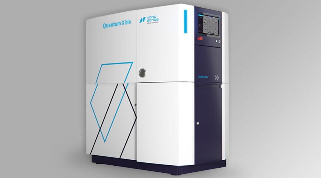
Larger biotechnology companies are also entering the fray. For example, BICO (formerly CELLINK) acquired Nanoscribe, signaling a strategic investment in ultra-high-resolution bioprinting for life-science and biomedical applications. Nanoscribe’s Quantum X platform extends two-photon polymerization beyond microfabrication into true 3D bioprinting, enabling the fabrication of cell-laden hydrogel structures with sub-micron precision. The system allows precise spatial placement of cells, extracellular matrix analogs, and microvascular features within complex 3D constructs, capabilities that are directly relevant to reproductive biology research, including engineered ovarian tissue models, endometrial interfaces, and advanced embryo culture environments.
The technological implications are significant: if additive manufacturing tools can improve embryo viability, reduce laboratory workload, and support more advanced screening workflows, such as high-throughput genetic analysis, they could materially improve IVF success rates and clinical efficiency. In practice, IVF clinics may increasingly deploy integrated AM-enabled solutions, ranging from microfluidic sperm sorters and embryo culture chips to safer cryopreservation systems and, eventually, bioprinted tissue constructs designed to support implantation and early pregnancy.
With rapid deployment of such technology, a light ethical framing is prudent. The same tools that allow us to culture and screen embryos with greater efficacy also inch us toward more controversial possibilities. For instance, polygenic embryo screening (enabled by fast on-chip DNA analysis) raises questions about selecting embryos for non-medical traits or the fairness of access to such enhancements. The use of bioprinted ovarian tissue or gametes intersects with debates on the extent of human germline intervention. Ensuring these advanced techniques are safe, validated, and equitably available will be a priority. Regulatory bodies will need to assess 3D-printed medical devices for reproductive use, but current studies showing biocompatibility and improved outcomes (e.g. the ICSI Pod system causing no harm to embryos while improving consistency) are encouraging.
From a societal perspective, surveys have indicated a general support for using technology to screen embryos for disease risk, but less so for trait selection. Thus, as engineers and clinicians innovate, ethicists and policymakers must keep pace to set appropriate guidelines.
The Research & Development Tax Credit
The now permanent Research and Development (R&D) Tax Credit is available for companies developing new or improved products, processes, and/or software. 3D printing can help boost a company’s R&D Tax Credits. Wages for technical employees creating, evaluating, and revising 3D printed prototypes are typically eligible expenses toward the R&D Tax Credit. Similarly, when used as a method of improving a process, time spent integrating 3D printing hardware and software can also be an eligible R&D expense. Lastly, when used for modeling and preproduction, the costs of filaments consumed during the development process may also be recovered.
Whether it is used for creating and testing prototypes or for final production, 3D printing is a great indicator that R&D Credit-eligible activities are taking place. Companies implementing this technology at any point should consider taking advantage of R&D Tax Credits.
Conclusion
Additive manufacturing is emerging as a transformative force in reproductive medicine. By enabling the fabrication of highly specialized microfluidic devices, biocompatible scaffolds, precision tools, and advanced cryopreservation systems, 3D printing addresses long-standing technical challenges in IVF and embryo screening. These technologies are progressing from experimental prototypes to clinically relevant tools, offering improvements in efficiency, safety, and reproducibility. As collaboration continues between engineers, clinicians, and researchers, additive manufacturing is poised to play a central role in the future of assisted reproduction, with tangible benefits for patients and laboratories alike.